
Chiari I malformation, CM1, is a frequent reason for neurosurgical consultation following MRI, often as an incidental finding. It occurs in both paediatric and adult neurosurgery, and the literature contains established recommendations, expert opinion and controversial techniques. This article synthesises the 2023 Congress of Neurological Surgeons, CNS, guideline, the 2022 international paediatric and adult consensus documents, and the Primer published in Nature Reviews Disease Primers in 2026.
A spectrum, not just a measurement
The classical definition is based on descent of the cerebellar tonsils through the foramen magnum. The 2026 Primer by Hale and colleagues emphasises that this anatomical criterion does not explain clinical variability. Some patients with minimal descent have severe symptoms and syringomyelia, while others with marked descent have no symptoms. The authors propose viewing CM1 as a spectrum of disturbances in the development and homeostasis of the cerebellum, posterior fossa, craniocervical junction and cerebrospinal fluid dynamics. In this model, tonsillar herniation is a geometric consequence rather than the primary process.
The practical message is straightforward: millimetres alone do not determine the indication.
Definitions and classification
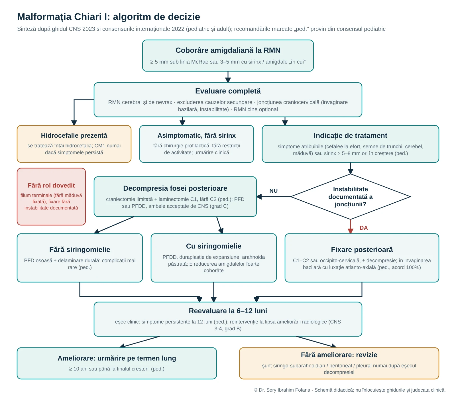
- Chiari I. The paediatric consensus defines CM1 as herniation of one or both cerebellar tonsils at least 5 mm below the McRae line. Descent of 3–5 mm is also accepted when syringomyelia or peg-like tonsils are present. Descent of 3–5 mm without these features is often described as borderline.
- Chiari 0. This remains controversial: syringomyelia with minimal or absent tonsillar herniation, below 3 mm, a small posterior fossa and an obliterated cisterna magna. Some authors regard it as a CM1 subtype, others as a distinct malformation.
- Chiari 1.5. Usually described as CM1 with descent of the brainstem, specifically the obex, below the foramen magnum. The paediatric consensus notes that it may coexist with basilar invagination and that only symptomatic cases should undergo surgery.
- Chiari II. Occurs with open spinal dysraphism, or myelomeningocele, and has its own management pathway linked to the spinal defect and hydrocephalus.
- Chiari III and IV. Rare forms in the classical classification: type III is associated with an occipital or upper cervical encephalocele, while the term type IV, used for cerebellar hypoplasia, is now considered outdated.
Which symptoms may be attributable to the malformation
The most characteristic presentation is occipital headache triggered by coughing, sneezing or straining, the Valsalva manoeuvre. Signs of brainstem, cerebellar or spinal cord dysfunction, otoneurological symptoms and scoliosis may occur, the latter particularly with syringomyelia.
The CNS guideline states that decompression may be performed in symptomatic patients to improve strain-related headache. Other symptoms respond more variably, so expectations must be discussed with the patient.
The paediatric consensus also states that:
- epilepsy does not improve through CM1 surgery;
- autism spectrum or behavioural disorders without clear Chiari symptoms are not an indication for surgery;
- tonsillar ectopia of even 20 mm or more does not by itself determine the need for intervention.
Assessment before deciding
- MRI of the brain and entire neuraxis. Syringomyelia must be excluded or characterised. In children, the consensus recommends neurological follow-up and neuraxis MRI until growth is complete.
- Exclude secondary causes. Hydrocephalus, space-occupying lesions and other causes of tonsillar descent must be excluded. If hydrocephalus is present, the paediatric consensus recommends treating it first; CM1 is addressed subsequently only if symptoms persist.
- Assess the craniocervical junction. Look for basilar invagination and atlanto-axial instability, using targeted imaging when suspected.
- Cine MRI, studying CSF flow. It may help but is not necessary in every case.
- Sleep or swallowing studies. These are not routinely recommended without corresponding symptoms. CNS considers the evidence insufficient for systematic use.
Surgical indications
- Asymptomatic CM1 without syringomyelia. Prophylactic surgery is not performed and activity restrictions are not recommended, according to the CNS grade C guideline and paediatric consensus. A small proportion may develop symptoms later, so clinical follow-up remains important.
- Asymptomatic CM1 with syringomyelia. In children, the consensus indicates surgery for a syrinx larger than 5–8 mm or a smaller but enlarging syrinx.
- Symptomatic CM1. Decompression is first-line treatment.
- Relatives. Routine screening of asymptomatic siblings or first-degree relatives is not recommended, CNS grade C.
Surgical options
1. Bony posterior fossa decompression (PFD)
Suboccipital craniectomy and C1 laminectomy without opening the dura. Dural splitting, separating the outer dural layer, is sometimes added. The paediatric consensus regards this as an option for symptomatic children without syringomyelia because of its low complication rate.
Two technical details from the paediatric consensus:
- decompression always includes C1 laminectomy and does not extend to C2 because of the risk of craniocervical instability;
- the craniectomy should not be wide, to avoid cerebellar ptosis.
2. Decompression with expansile duraplasty (PFDD)
The dura is opened and an autologous or heterologous graft enlarges the space at the junction and restores the cisterna magna. This is the posterior fossa ‘expansion’. The paediatric consensus recommends preserving the arachnoid when arachnoiditis is absent to reduce CSF leakage and late scarring.
CNS recommendation 3-1, grade C, accepts both PFD and PFDD as first-line treatment because no class I evidence favours either. The reviewed evidence nevertheless describes a clear trade-off:
- syringomyelia: most studies report better resolution after PFDD. A study published after the guideline's search period, Akbari 2022, found syrinx reduction in 47.7% with PFDD versus 26.9% after PFD;
- complications: pseudomeningocele and CSF leakage are more common after PFDD. In a class II study, Pisapia 2016, opening the dura was associated with pseudomeningocele in 18% and chemical meningitis in 10%, versus 0% after PFD, without a difference in reoperation rate;
- reoperations: these were more frequent after PFD, but the difference was not statistically significant.
The paediatric consensus concludes that bony decompression with duraplasty is preferable when syringomyelia is present.
3. Cerebellar tonsillar reduction
Tonsillar coagulation or subpial resection may be performed to improve the syrinx or symptoms, CNS recommendation 3-2, grade C. Evidence does not establish that it is needed for every patient subgroup, and some studies reported more complications. The paediatric consensus reserves it for very low-lying tonsils and recurrent or residual syringomyelia.
4. Craniocervical fixation (C1–C2 or occipitocervical)
This is the greatest area of controversy. Goel proposed ‘central’ atlanto-axial instability, sometimes apparent only intraoperatively, as a common cause of CM1, basilar invagination and syringomyelia. He recommends C1–C2 fixation without decompression. A critical literature review by Wagner and colleagues in 2020 found low-level evidence and insufficient support for the hypothesis. The authors regarded abandoning established procedures in favour of a single approach as unjustified.
The paediatric consensus states that:
- for basilar invagination with atlanto-axial dislocation, posterior fixation is preferred, with 100% agreement;
- for basilar invagination without atlanto-axial instability, posterior reduction is attempted first, with anterior decompression added if this fails;
- fixation is not indicated in CM1 without documented craniocervical instability, with 90.5% agreement.
5. Filum terminale sectioning
The hypothesis of ‘caudal traction’ from an occult tethered cord led to proposals for filum terminale sectioning as CM1 treatment. A systematic review by Milano and colleagues in 2020 found only two small series from the same group, with low methodological quality and level 4 evidence. Its conclusion was that there is no scientific support for the procedure in CM1 without tethered cord syndrome and that it should be considered experimental.
The paediatric consensus is equally clear, with 100% agreement:
- intradural filum terminale sectioning is recommended only to treat tethered cord syndrome;
- it has no role in CM1;
- extradural sectioning is not recommended.
6. Direct treatment of syringomyelia
Syringosubarachnoid, syringoperitoneal or syringopleural shunts are considered only after failure of treatment directed at the cause, namely decompression.
7. Intraoperative neuromonitoring
It may be used, CNS recommendation 3-3, grade C, with an individual decision. The evidence reviewed by the guideline did not clearly demonstrate benefit or harm from neuromonitoring.
Cerebellar ptosis and posterior fossa reconstruction
An excessively wide suboccipital craniectomy may allow cerebellar descent with recurrence of symptoms. Symptomatic cerebellar ptosis after wide decompression is reported in the literature and may require posterior fossa reconstruction. The best prevention remains a limited craniectomy, as recommended by the paediatric consensus.
Postoperative course and failure
- Syringomyelia. Improvement is slow: most cases improve over 6–12 months, with a median of eight months to 50% regression, Chotai 2020, cited by the CNS guideline.
- Reoperation. Further intervention may be performed at 6–12 months in patients without radiological improvement, CNS recommendation 3-4, grade B.
- Clinical failure. In children, the consensus defines this as symptoms persisting at 12 months. Reoperation is indicated for persistent symptoms with unchanged MRI at 6–12 months.
- Follow-up. After successful surgery, the paediatric consensus recommends follow-up for at least 10 years or until growth is complete.
Key messages
- The indication for surgery is based on attributable symptoms and syringomyelia, not millimetres.
- Treat hydrocephalus first.
- For a symptomatic child without a syrinx, bony decompression with C1 laminectomy is a valid option. When a syrinx is present, expansile duraplasty is preferable.
- Limit the craniectomy and preserve C2.
- Craniocervical fixation is performed only for documented instability or basilar invagination with atlanto-axial dislocation.
- Filum terminale sectioning has no role in CM1 without tethered cord syndrome.
- Assess failure at 6–12 months; syrinx shunting follows only after failure of treatment directed at the cause.
Recommendations marked ‘paediatric consensus’ come from the document for children. For adults, consult the 2023 CNS guideline, applicable to both age groups, and the international adult consensus, which contains 63 statements on definition, planning, surgery and isolated syringomyelia.