
Back pain radiating down a leg is one of the most common reasons for seeing a neurosurgeon. A lumbar disc herniation is often the cause. The diagnosis can sound worrying, but most cases improve without surgery. It is important to know which signs require prompt assessment and when discussing an operation makes sense.
What an intervertebral disc is
Intervertebral discs lie between the vertebrae and cushion spinal movement. Each disc has two parts: a tough outer fibrous ring, the annulus fibrosus, and a jelly-like central core, the nucleus pulposus.
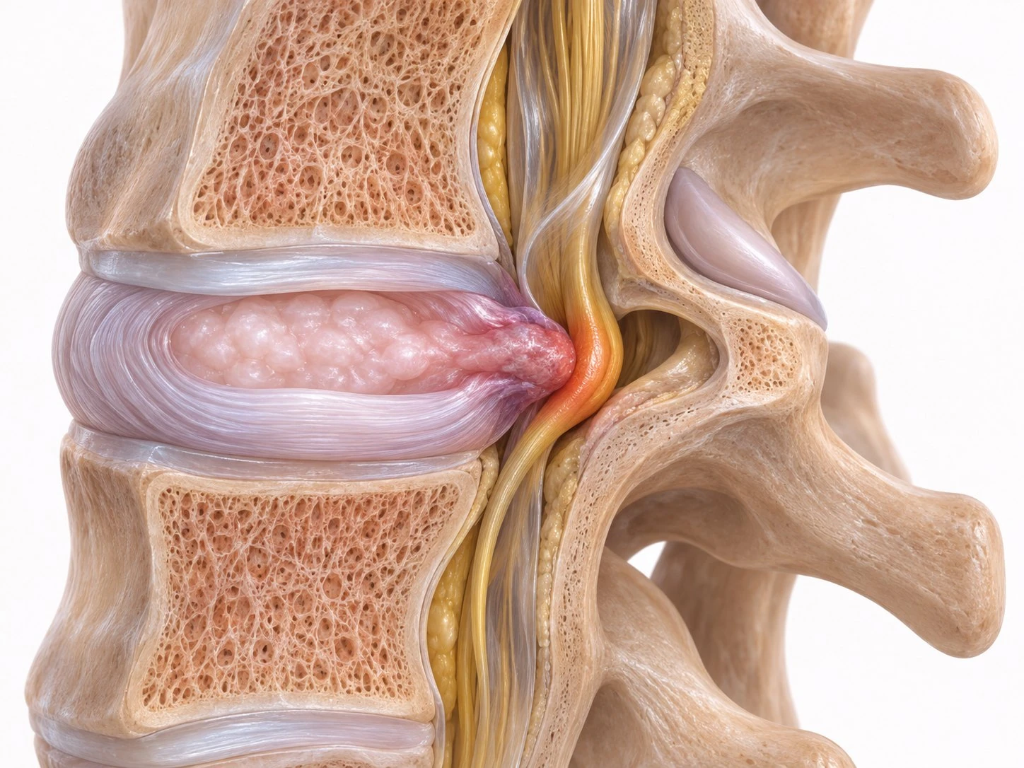
Disc herniation occurs when part of the nucleus pulposus passes through a tear in the annulus and extends beyond the disc's normal space. In the lumbar spine, the two lowest levels, L4–L5 and L5–S1, are most often affected.
Why the leg hurts
The disc fragment may press on and irritate a nerve root. Local inflammation also matters alongside mechanical compression. The result is pain following the nerve's course, usually down the back or side of the thigh and calf into the foot. This is called sciatica or, medically, lumbar radiculopathy.
Alongside pain, there may be:
- numbness or tingling in a particular part of the leg;
- weakness in certain muscles, such as difficulty walking on tiptoes or heels;
- reduced tendon reflexes detected on neurological examination.
What MRI shows and what it does not
Magnetic resonance imaging, or MRI, shows discs and nerve roots best. However, the images must be interpreted alongside the symptoms and clinical examination.
Degenerative disc changes are very common even in people without pain. In a large review of studies in asymptomatic people, disc protrusion was present in approximately 29% of 20-year-olds and 43% of 80-year-olds, while disc bulging was present in 30% and 84%, respectively. An MRI report describing ‘protrusions’ or ‘bulges’ therefore does not automatically explain pain.
For the same reason, the UK NICE guideline advises against routinely offering imaging outside specialist services for low back pain with or without sciatica. MRI is useful when the result could change management, for example when surgery is being considered or warning signs are present.
The usual course
For many patients, pain gradually decreases in the weeks after onset. The herniated fragment may shrink or disappear spontaneously as the body resorbs it.
A meta-analysis published in 2024 combining 31 studies and more than 2,200 patients treated without surgery found spontaneous resorption in approximately 70% of cases. Regression was more frequent in larger herniations:
- around 88% of sequestrated fragments, completely detached from the disc;
- around 67% of extrusions;
- around 38% of protrusions;
- around 13% of disc bulges.
Most resorption occurred within the first six months. These figures cannot predict an individual patient's course, but they help explain why treatment usually begins without surgery when warning signs are absent.
Non-surgical treatment
Initial treatment aims to control pain and maintain activity as close to normal as possible. Prolonged bed rest is not recommended. The plan is individual and may include:
- information and a gradual return to daily activities;
- exercise and a rehabilitation programme guided by a physiotherapist;
- pain medication selected and adjusted by a doctor, considering benefits and risks;
- in certain situations, injection procedures discussed with a specialist.
When surgery is discussed
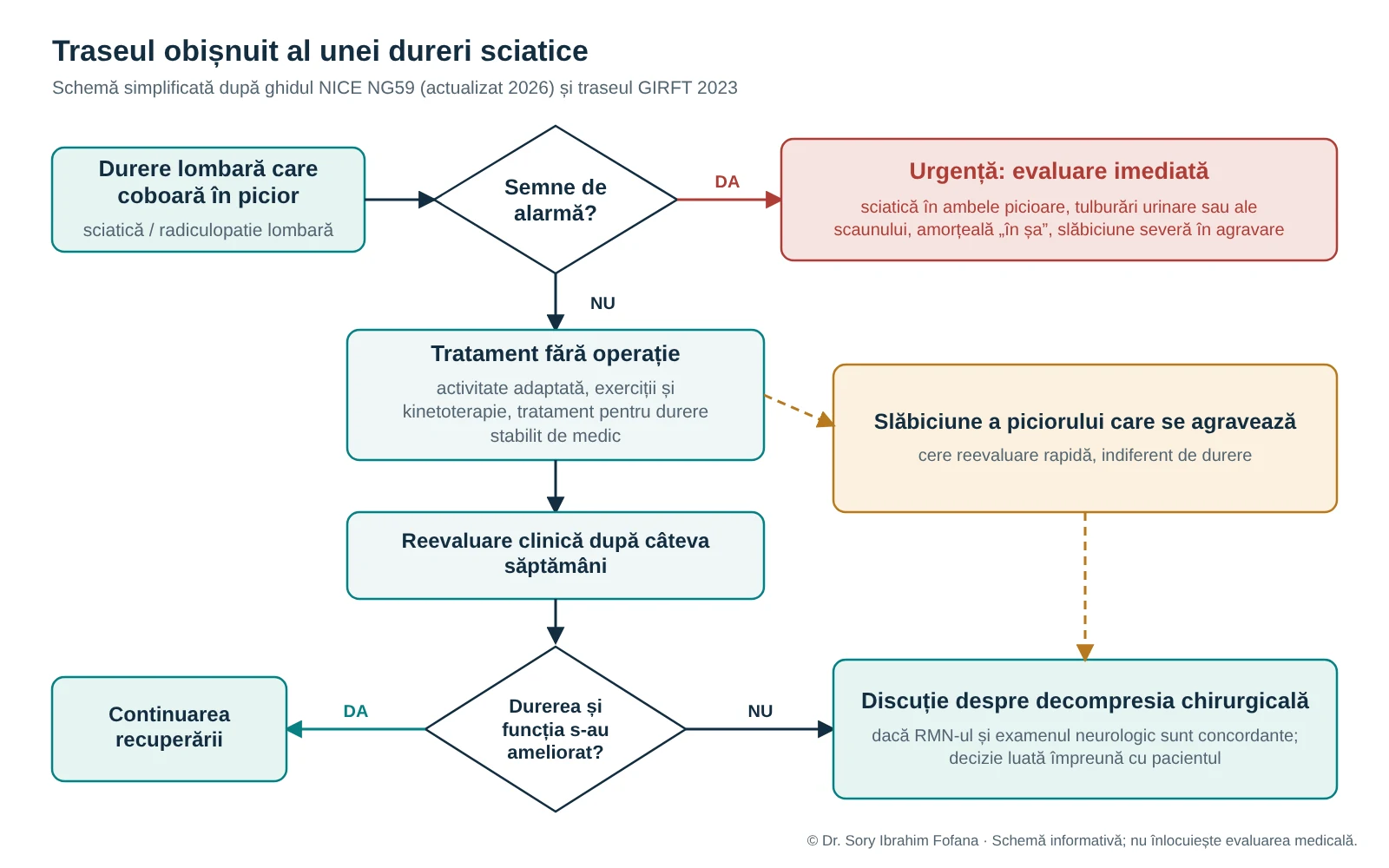
NICE recommends considering surgical decompression when non-surgical treatment has not improved pain or function and MRI findings match the symptoms. Agreement between the clinical examination and imaging is essential: surgery treats a patient with a defined problem, not an image.
A randomised trial published in the New England Journal of Medicine in 2020 included patients with sciatica lasting 4–12 months caused by an L4–L5 or L5–S1 herniation. At six months, leg pain was significantly lower after microdiscectomy than after non-surgical treatment, averaging 2.8 versus 5.2 on a 0–10 scale, with similar findings at one year. Around one third of patients initially treated without surgery later underwent an operation.
For pain persisting for months, surgery is therefore a well-supported option. The decision is made together with the patient, considering:
- the intensity and duration of pain;
- the impact on daily life and work;
- the presence and progression of weakness;
- agreement between neurological examination and MRI findings;
- the patient's preferences following a clear discussion of benefits and risks.
Worsening weakness requires prompt reassessment even if the pain has improved.
What microdiscectomy involves
The usual operation for lumbar disc herniation is microdiscectomy. Through a small incision and using optical magnification, the surgeon removes the disc fragment compressing the nerve root while preserving the structures that provide spinal stability. The main aim is to relieve leg pain. The benefit for isolated back pain may differ and is discussed separately before surgery.
Warning signs: when to go to hospital urgently
Rarely, a large disc herniation can compress several nerve roots at the bottom of the spinal canal. This is called cauda equina syndrome and requires emergency assessment. Go immediately to an emergency department if any of the following occurs:
- sciatic pain in both legs;
- severe or worsening weakness in both legs;
- difficulty starting to urinate or altered sensation when passing urine;
- loss of the sensation of rectal fullness;
- numbness between the legs, in the perineum or genital area, known as saddle numbness;
- new sexual dysfunction;
- loss of bladder or bowel control.
Do not wait for a routine appointment in these situations.
Useful questions before an operation
- What is the exact diagnosis and which spinal level is involved?
- Which symptom is most likely to improve after surgery?
- What alternatives are available and how long is it reasonable to try them?
- Which risks are relevant to my case?
- What will recovery involve and when can I return to normal activities?
What to bring to your consultation
The consultation is more useful when the doctor has all the information:
- the CD or DVD with the original MRI images in DICOM format, not only the written report;
- investigation reports and, if available, older studies for comparison;
- medical letters and a list of previous treatments, including physiotherapy;
- a brief note of onset, the course of pain and any new symptom.
Frequently asked questions
Can a disc herniation resorb without surgery?
Yes. The herniated fragment may shrink or disappear spontaneously. Regression is more common in larger extruded or sequestrated herniations, as shown by the imaging follow-up studies cited above.
Should I stay in bed until the pain passes?
No. Prolonged bed rest is not recommended. Guidelines encourage continuing usual activities within pain limits, together with adapted exercise.
I have a disc herniation on MRI but no leg pain. Does it need treatment?
An image without corresponding symptoms is not by itself an indication for treatment. Disc changes are common in people without pain, and the decision always follows clinical examination.