
Hydrocephalus is a condition in which the fluid surrounding and protecting the brain builds up excessively. In children it may develop before birth, during the first months of life or later. It is treatable but requires an accurate diagnosis, careful follow-up and sometimes neurosurgery. This article briefly explains what parents need to know.
What cerebrospinal fluid is
Inside the brain are cavities called ventricles. Cerebrospinal fluid (CSF) is continuously produced here. This clear fluid surrounds the brain and spinal cord, provides mechanical protection and helps maintain a stable environment for nervous tissue.
CSF normally flows from the lateral ventricles to the third ventricle, then through a narrow channel, the Sylvian aqueduct, to the fourth ventricle. It then enters the spaces around the brain and spinal cord, where it is absorbed into the venous circulation. Production and absorption are in balance.

Hydrocephalus develops when this balance is disturbed: fluid circulation is blocked or absorption is insufficient. CSF accumulates, the ventricles enlarge and intracranial pressure may rise.
Why it develops
Causes vary from one child to another:
- congenital causes, present from birth, such as narrowing of the Sylvian aqueduct or other malformations;
- prematurity, particularly after a brain haemorrhage in a newborn;
- infections such as meningitis, which can affect fluid absorption;
- brain tumours that may obstruct CSF circulation;
- in many cases, the exact cause cannot be identified.
Signs parents may notice
In infants, the skull bones have not yet fused, so increasing head size is often the first sign. Signs to discuss with a doctor include:
- head growth faster than expected on repeated head circumference measurements;
- a bulging or tense fontanelle, the soft area on top of the head;
- thin, shiny scalp with easily visible veins;
- downward-directed gaze (‘sunsetting eyes’);
- vomiting, irritable crying or refusal to feed;
- unusual drowsiness or reduced responsiveness;
- pallor.
In older children, increasing head circumference is less apparent, and symptoms may include:
- headaches, particularly in the morning;
- vomiting;
- tiredness and drowsiness;
- behavioural changes or declining school performance;
- blurred or double vision, and balance or walking difficulties.
None of these signs automatically means hydrocephalus, but a combination of signs or rapid worsening requires prompt medical assessment.
How it is diagnosed
The paediatrician measures head circumference at each check-up and compares it with growth charts. If hydrocephalus is suspected, imaging investigations are recommended:
- cranial ultrasound through an open fontanelle in infants: it is quick, painless and does not use radiation;
- brain MRI, which shows the ventricles, any obstruction and its cause in greatest detail;
- brain CT, mainly used in emergencies because it can be performed very quickly.
Alongside imaging, the clinical examination, the child's development and changes over time matter. Sometimes the ventricles are slightly enlarged but stable and the child is developing normally. In such cases, follow-up alone may be recommended.
Treatment options
When hydrocephalus causes symptoms or progresses, treatment is usually neurosurgical. There are two main types of procedure.
Ventriculo-peritoneal shunt
A shunt is a thin silicone tube fitted with a valve. One end is placed in a brain ventricle and the other runs under the skin to the abdominal cavity, the peritoneum, where excess fluid is absorbed. Less commonly, fluid is drained elsewhere. The valve controls how much fluid drains.
A shunt works for many children, but it may need revision during their lifetime, for example if it becomes blocked or infected. The Congress of Neurological Surgeons guideline, updated in 2020, recommends antibiotic-impregnated catheters in children to reduce the risk of shunt infection.
Endoscopic third ventriculostomy
In certain forms of hydrocephalus, particularly when fluid is blocked at a single point, a new circulation pathway can be created. The neurosurgeon introduces an endoscope into the ventricles and makes a small opening in the floor of the third ventricle. Fluid bypasses the obstruction and reaches spaces where it can be absorbed. The advantage is that no implanted device remains.
Not every child is a suitable candidate. Success depends mainly on the child's age, the cause of hydrocephalus and any previous shunt. These factors are combined in a prediction tool, the ETV Success Score, used when choosing treatment. A prospective multicentre Hydrocephalus Clinical Research Network study published in 2024 included 761 children treated using this technique: it was successful at six months in around three quarters of them, 76%, and the prediction score retained its value.
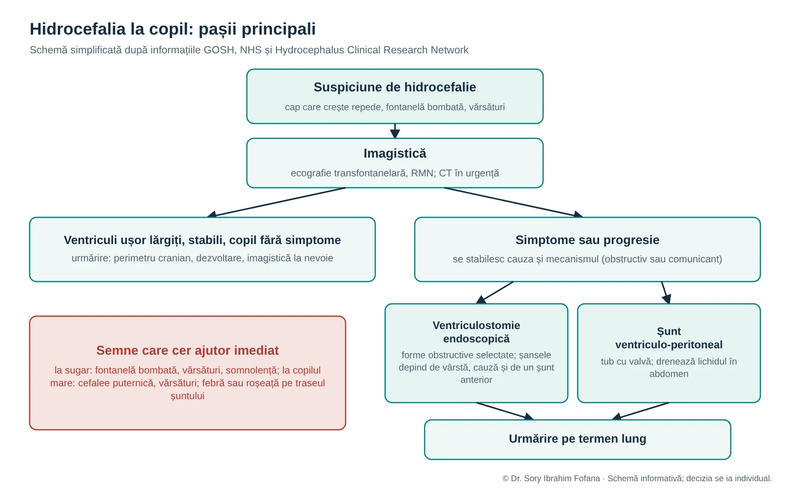
The choice between these approaches is individual, following review of the images and discussion with the family.
After surgery: signs requiring immediate help
After a shunt is inserted, parents need to know the signs of malfunction and infection. Shunt blockage can be serious and may require emergency surgery.
Contact the medical team immediately or attend an emergency department if you notice:
- in an infant: a bulging fontanelle, increasing head size, sunsetting eyes, vomiting, drowsiness or pallor;
- in an older child: severe headache, vomiting or unusual drowsiness;
- fever, redness or tenderness along the shunt, neck stiffness or abdominal pain;
- any sudden change in the child's condition that concerns you.
Shunt infection occurs most often in the first months after surgery. Signs of obstruction may develop at any time.
Long-term follow-up
Many children with treated hydrocephalus attend school, take part in sport and lead active lives. A shunt has no fixed ‘expiry date’: it is revised when a problem occurs. Follow-up is also needed after endoscopic ventriculostomy because the opening may close over time. Regular follow-up includes neurosurgical reviews, developmental assessment and imaging when needed. It is helpful for the family to keep all investigations and details of the shunt or procedure for future consultations.