The word ‘tumour’ in an MRI report almost always causes concern. With meningioma, it helps to know from the outset that most are benign and slow-growing. Many do not need an immediate operation. The right decision nevertheless depends on several factors, explained below.
What a meningioma is
The brain and spinal cord are covered by three membranes called the meninges. A meningioma arises from cells in these membranes, most often cells of the layer called the arachnoid.
Because it arises from the covering, a meningioma grows outside brain tissue. As it enlarges, it may displace and compress the brain, nerves or nearby vessels. This distinguishes it from tumours arising within the brain itself, such as gliomas, which behave differently and need different treatment.
It is also helpful to distinguish primary intracranial tumours, arising from the brain or surrounding structures, from metastases, originating from cancer in another organ. Meningioma belongs to the first category.
How common it is and who is affected
Meningioma is the most common primary intracranial tumour, meaning a tumour that starts within the skull rather than spreading from another organ. The US CBTRUS registry report published in 2025 indicates that meningiomas account for approximately 43% of primary brain and central nervous system tumours.
Other important facts:
- 80–85% of meningiomas are benign, WHO grade 1;
- around 15–18% are atypical, grade 2, and 1–3% are malignant, grade 3;
- they are 2–3 times more common in women than men;
- the median age at diagnosis is approximately 65 years.
How it is found
Some meningiomas cause symptoms by compressing nearby structures. Symptoms depend on location and may include:
- headaches;
- seizures;
- visual disturbance;
- changes in behaviour or memory;
- weakness in a limb.
Many meningiomas are discovered incidentally on MRI or CT performed for another reason. These are called incidental meningiomas. Autopsy studies suggest that up to 2.8% of people have a meningioma, most of them small and without consequences.
How it is diagnosed
Contrast-enhanced brain MRI is the reference investigation for diagnosis and follow-up. It shows the tumour's size, location, relationship to the brain, nerves and vessels, and any surrounding oedema.
CT is particularly useful for assessing skull bone next to the tumour.
The definitive diagnosis and tumour grade are established by histopathological examination after surgery or biopsy. The grade depends on the cells' microscopic appearance, division rate, brain invasion and, more recently, certain tumour genetic changes.
Treatment options
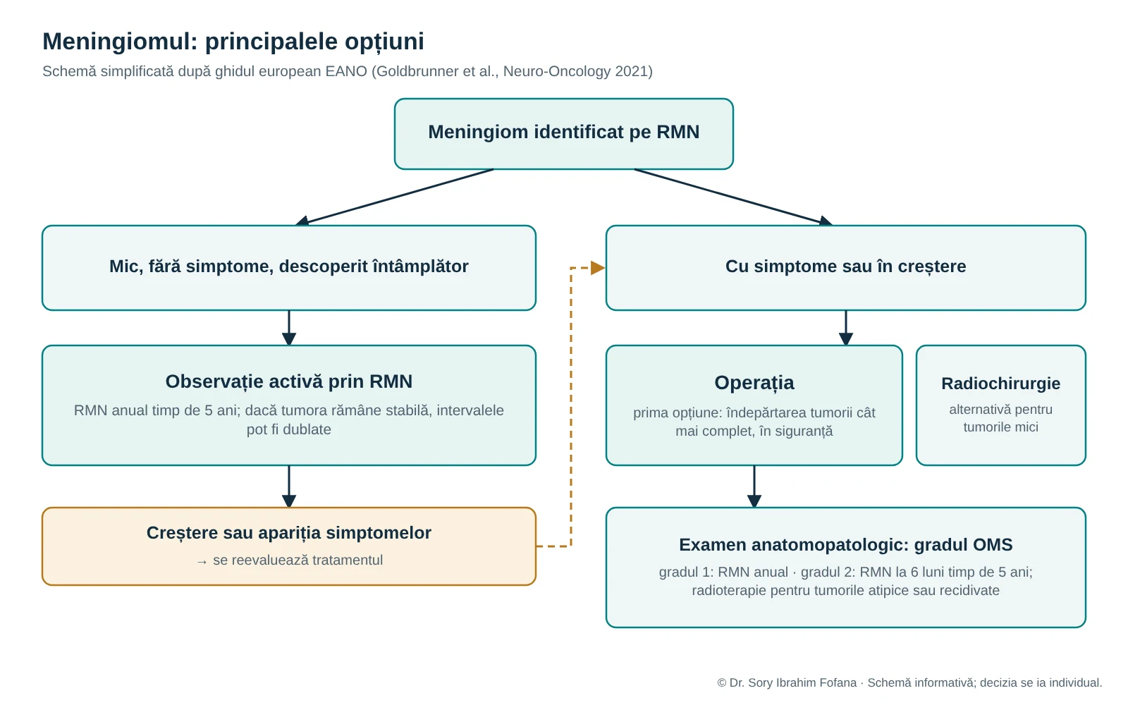
The European Association of Neuro-Oncology, EANO, guideline describes three main approaches.
MRI monitoring
For an incidentally discovered meningioma without symptoms, observation is the first option. The guideline recommends annual MRI for five years for suspected or confirmed grade 1 meningiomas. If the tumour remains stable, intervals can then be doubled.
Monitoring does not mean ‘doing nothing’. It is an active strategy that avoids unnecessary surgery and allows intervention at an appropriate time if the tumour grows or symptoms develop.
Surgery
For growing or symptomatic meningiomas, surgery is the first option. The aim is to remove as much tumour as safely possible. Sometimes the location, such as proximity to important nerves or vessels, leads to a planned partial removal followed by observation or other treatments.
Radiosurgery and radiotherapy
Stereotactic radiosurgery delivers a precisely targeted radiation dose, usually in one session. In selected cases involving small tumours, it may be an alternative to surgery. Fractionated radiotherapy, delivered over several sessions, is used particularly after surgery for atypical or recurrent tumours.
What influences the decision
There is no single treatment for every meningioma. Doctor and patient consider together:
- the tumour's size and location;
- whether it is growing and how quickly;
- symptoms;
- age and general health;
- the patient's preferences and goals.
After surgery, follow-up frequency depends on tumour grade. For grade 2 meningiomas, for example, EANO recommends MRI every six months for the first five years, then annually.
When not to wait for a routine appointment
A new seizure, sudden weakness, a new speech disturbance, confusion or impaired consciousness requires emergency medical assessment. Call 112 or attend an emergency department.
Useful questions for the consultation
- How large is the tumour and exactly where is it?
- Has it grown compared with previous investigations?
- Does the tumour explain my symptoms?
- What options do I have: monitoring, surgery or radiosurgery?
- What are the benefits and risks of each option in my case?
- What will follow-up involve over the coming years?
To help answer these questions accurately, bring the original MRI images in DICOM format, the reports and older investigations for comparison.