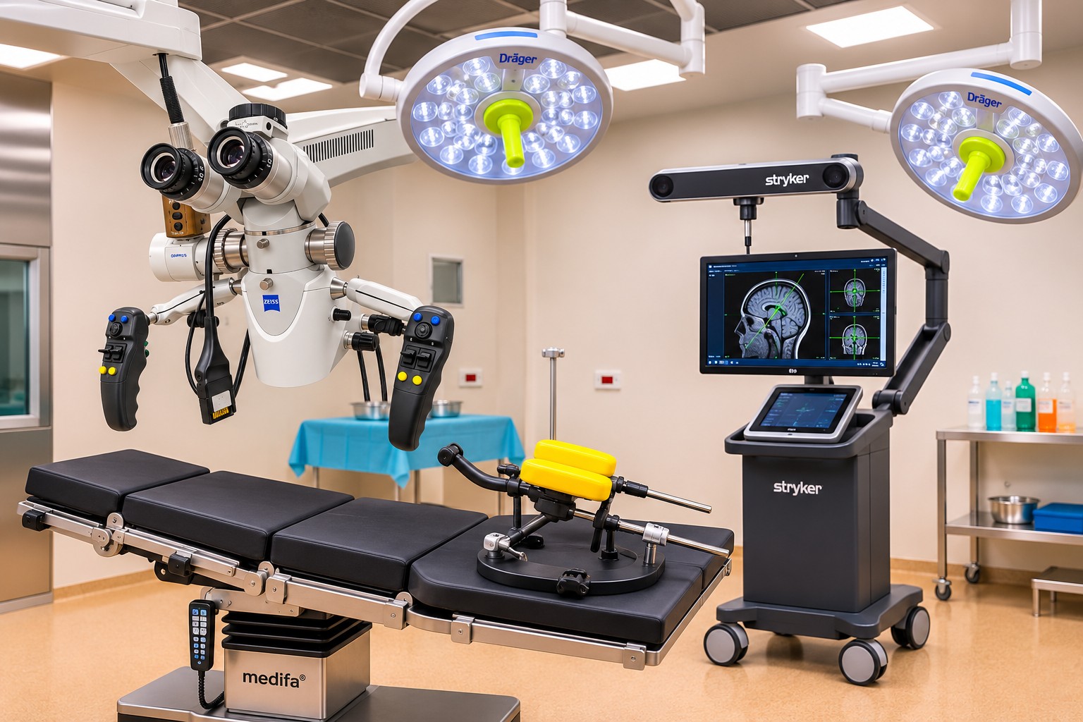
Someone entering a neurosurgical operating theatre for the first time usually notices the equipment: the microscope suspended over the table, monitors, fine instruments and navigation system. For the neurosurgical team, however, the operation starts earlier and somewhere much quieter, reviewing the patient's images and trying to understand what lies beyond what the screen shows.
Every person's anatomy is different. A tumour may lie against an important vessel, a cyst may block cerebrospinal fluid circulation, or a herniated disc may press on a nerve root within a few millimetres of space. Reaching the lesion is rarely the only challenge. The difficult part is treating it while protecting what still functions well: movement, sensation, speech and vision. This is where technology provides meaningful support for clinical experience.
Imaging, DICOM and planning
The starting point is usually MRI or computed tomography. An MRI examination is not a photograph but hundreds of images grouped into sequences that show the same tissues in different ways. We review them in several planes and compare them with older studies. The radiologist's report matters, but a surgical decision also requires the original images. Patients are therefore asked to bring the investigation CD or access link, not only the written result. The original images are retained in DICOM format together with all the examination sequences.
Planning software can combine these sections into a three-dimensional volume. The volume consists of very small points called voxels, similar to photographic pixels but with depth. They can be used to reconstruct the outline of a lesion, blood vessels, ventricles or brain surface. Sometimes two investigations, such as MRI and CT, are overlaid through image fusion to show the relationship between brain, bone and vessels in one view. In selected cases, tractography estimates the course of nerve fibre bundles, for example those controlling movement. It is an estimate rather than an exact map, but may change the direction from which a lesion is approached.
The reconstructions may look impressive, but they are used for specific questions. Which route is safer? Which structures must be protected? How large should the opening be? What if the findings during surgery differ from expectations? A well-prepared operation has several possible approaches considered before the first incision.
Neuronavigation and brain shift
In the operating theatre, the plan meets reality. Neuronavigation works a little like GPS: it links preoperative images to the patient's position on the table and shows an instrument tip's position relative to that map. It is especially helpful when disease has distorted normal landmarks. But it has a limitation familiar to every neurosurgeon. After the skull is opened, as fluid drains or tumour tissue is removed, the brain may shift by several millimetres, sometimes more than a centimetre. The map remains the preoperative one, while the brain is no longer in exactly the same position. The screen is therefore continually checked against what the surgeon sees and feels through the instruments.
Microscopy and neuroendoscopy
The operating microscope provides light and magnification where structures are very delicate. Near a nerve or small vessel, the difference between a safe and risky manoeuvre may be a fraction of a millimetre.
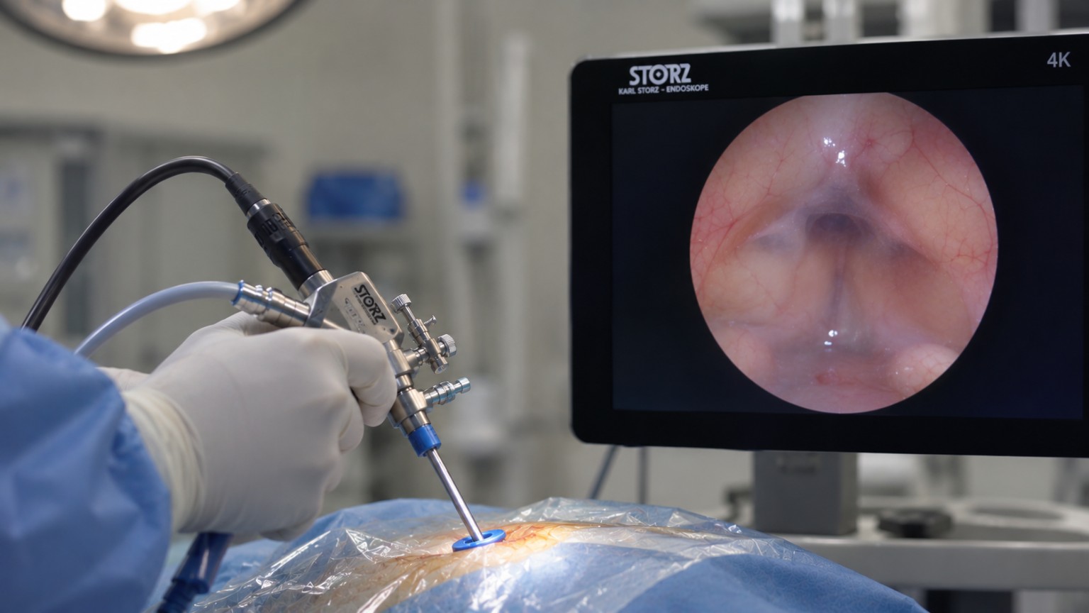
Neuroendoscopy offers a different possibility: a slender camera introduced through a small opening, allowing work in narrow spaces such as the brain's ventricles in some forms of hydrocephalus. A small approach is not an aim in itself. It must be the right approach for the condition, and sometimes that means a wider one.
Intraoperative assessment
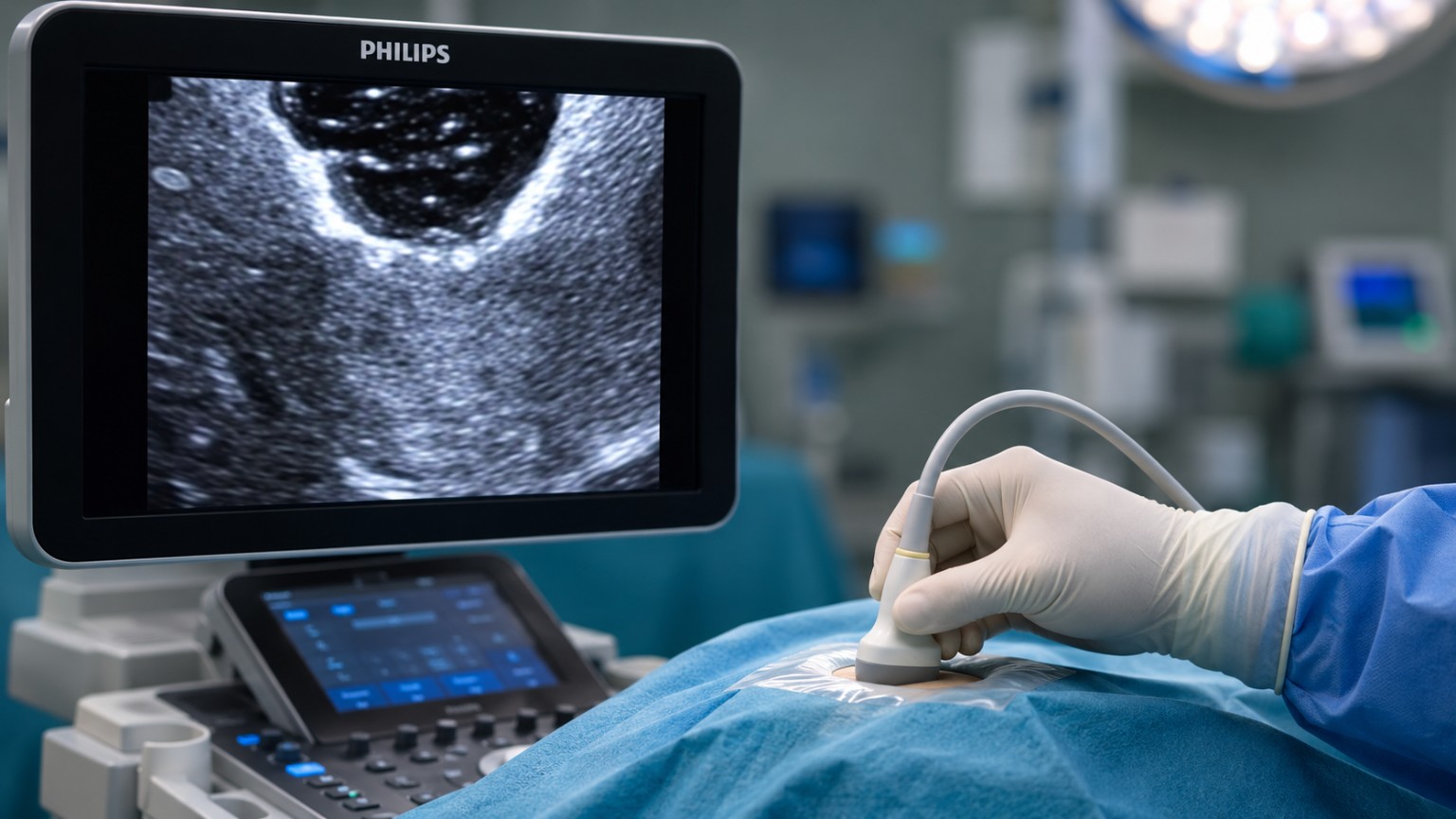
Other technologies provide information during the operation itself. Intraoperative ultrasound offers an up-to-date view of tissues and can partly account for the shift described above.
When indicated, neuromonitoring follows electrical signals in selected neural pathways and alerts the team if a manoeuvre begins to affect them. Fluorescence is used for some brain tumours: a substance given before surgery causes tumour tissue to fluoresce under a special microscope light. Each technique addresses a different question, and not all are needed in every operation.
Digital image processing and artificial intelligence
Digital image processing has advanced considerably in recent years. Some programs automatically outline structures, take measurements or compare studies performed months apart. Artificial intelligence tools can shorten this work, but their output must be checked by a doctor. An algorithm does not know what brought the patient to the consultation, which symptom frightens them, or what returning to work, driving or holding their child means to them.
Technology within clinical decision-making
When discussing an operation with a patient, we therefore try to explain less about the equipment and more about why surgery is recommended. Sometimes an operation is the answer. At other times, monitoring a lesion, obtaining another investigation or discussing the case with colleagues in other specialties is more appropriate. When surgery is considered unnecessary, that is explained just as clearly. For the team, precision begins with the choice of treatment.
Good equipment helps us see more, orient ourselves and work with more information. Its value depends on the team using it: the questions they ask, how they interpret the answers and when they recognise the need to change the plan. In neurosurgery, progress ultimately shows in the decisions made for each patient.