Finding an intracranial lesion on MRI is understandably unsettling. However, ‘brain tumour’ describes very different situations. Some lesions are benign and slow-growing, others malignant. Some arise within the brain, others in surrounding structures, and sometimes an image only resembles a tumour. They do not all need the same management or immediate surgery.
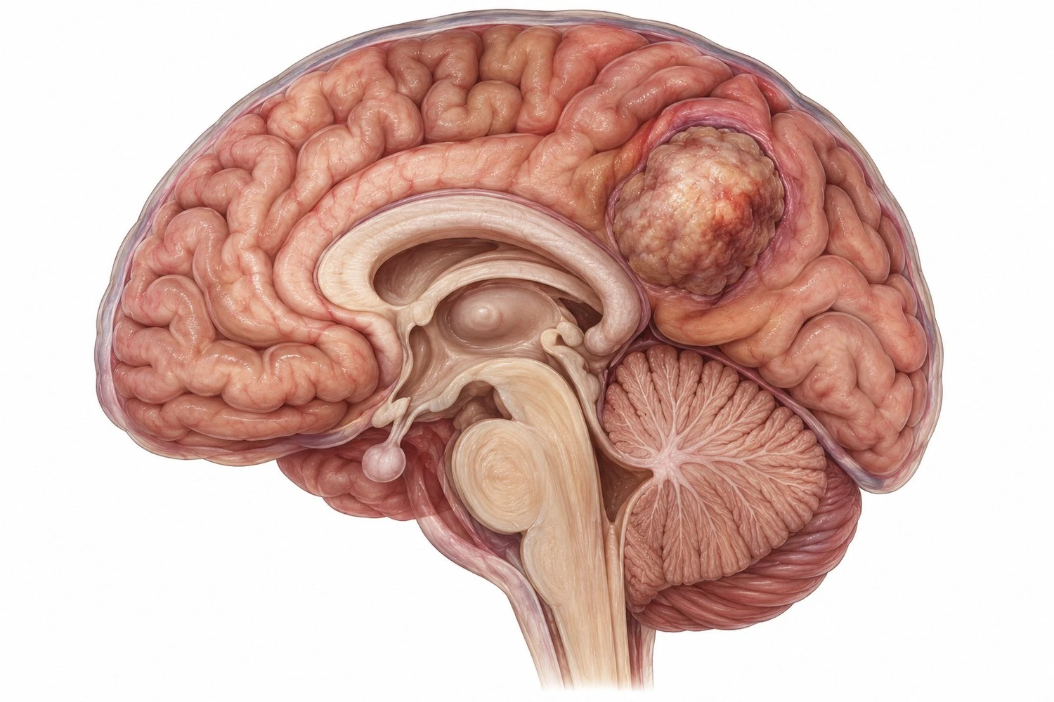
An image is not yet a complete diagnosis
The first step is to understand the images accurately alongside the symptoms and neurological examination. Ideally, the complete DICOM study is reviewed, rather than only the written conclusion or a few photographs.
Intracranial tumours fall into two broad categories:
- primary tumours, arising from the brain or surrounding structures, such as glioma or meningioma;
- brain metastases, originating from cancer in another organ, such as the lung or breast.
What the consultation clarifies
The consultation considers why the MRI was performed, current symptoms, their progression and associated illnesses. Important imaging features include:
- the lesion's location and size;
- its relationship to areas controlling movement, language or vision;
- surrounding oedema and mass effect on neighbouring structures;
- its appearance on different MRI sequences and, where available, comparison with older investigations.
The UK NICE guideline recommends MRI as the initial investigation for suspected glioma or meningioma when there are no contraindications. Additional sequences, other investigations or assessment by other specialties are sometimes needed.
Observation, biopsy or surgery?
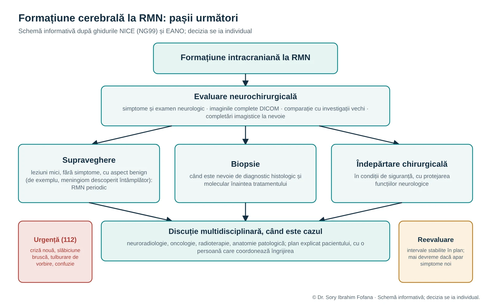
There is no single answer for every tumour. Depending on the situation, the main options are:
- Clinical and imaging surveillance, for example for a small, incidentally discovered meningioma without symptoms. EANO guidance recommends observation with periodic MRI in this setting.
- Biopsy when a histological and molecular diagnosis is needed before choosing treatment.
- Surgical removal. The aim is not simply to ‘remove everything’, but to treat the lesion safely while protecting neurological function as well as possible.
The decision considers the likely lesion type, progression, age, general health, the patient's preferences and possible complementary treatments such as radiotherapy or oncological therapy.
Why the multidisciplinary team matters
In oncological cases, decisions are usually made with other specialties: neuroradiology, oncology, radiotherapy and pathology. NICE recommends discussing suspected glioma with a specialist multidisciplinary team from the first radiological diagnosis. It also recommends a designated professional to coordinate each patient's care and information that is realistic, empathetic and provided at the right time.
When not to wait for a routine appointment
Attend an emergency department immediately or call 112 if any of the following occurs:
- a new seizure;
- sudden weakness on one side of the body;
- a new speech disturbance;
- marked confusion or unusual drowsiness;
- rapid neurological deterioration.
A scheduled consultation does not replace emergency care.
What to bring to the assessment
- complete MRI or CT studies on CD, DVD or USB in DICOM format;
- radiology reports;
- previous investigations for comparison;
- a list of treatments and associated illnesses;
- a brief timeline of symptoms.
If surgery has already been recommended, write down what you would like to clarify: the diagnosis, urgency, alternatives, benefits, risks and subsequent follow-up.
Useful questions for the consultation
- What type of lesion does this appear to be, and how certain is the diagnosis?
- Does it explain my symptoms?
- Is treatment needed now, or is monitoring reasonable?
- What options do I have, and what are their benefits and risks?
- Who will coordinate the next steps, and when is the next assessment?